How the Fruit Fly Brain Map Was Made: 7,062 Slices, 21 Million Images, 3 Million Edits
How the Fruit Fly Brain Map Was Made: 7,062 Slices, 21 Million Images, 3 Million Edits
In October 2024 the FlyWire Consortium published the first complete wiring diagram of an adult fruit fly brain in Nature: 139,255 neurons and 54.5 million synapses, with every neuron's partners and synapse counts mapped.
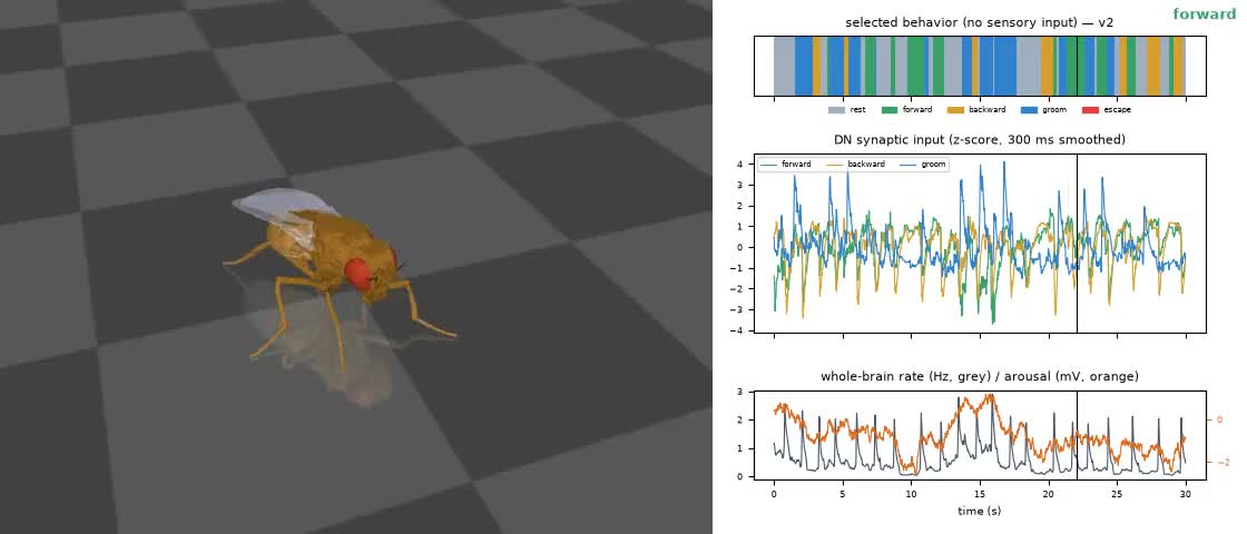
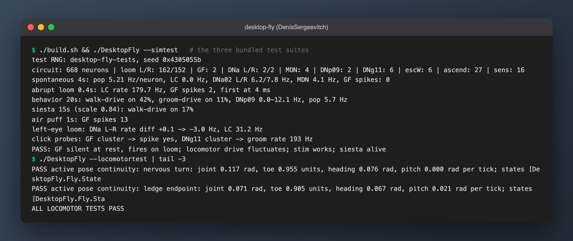
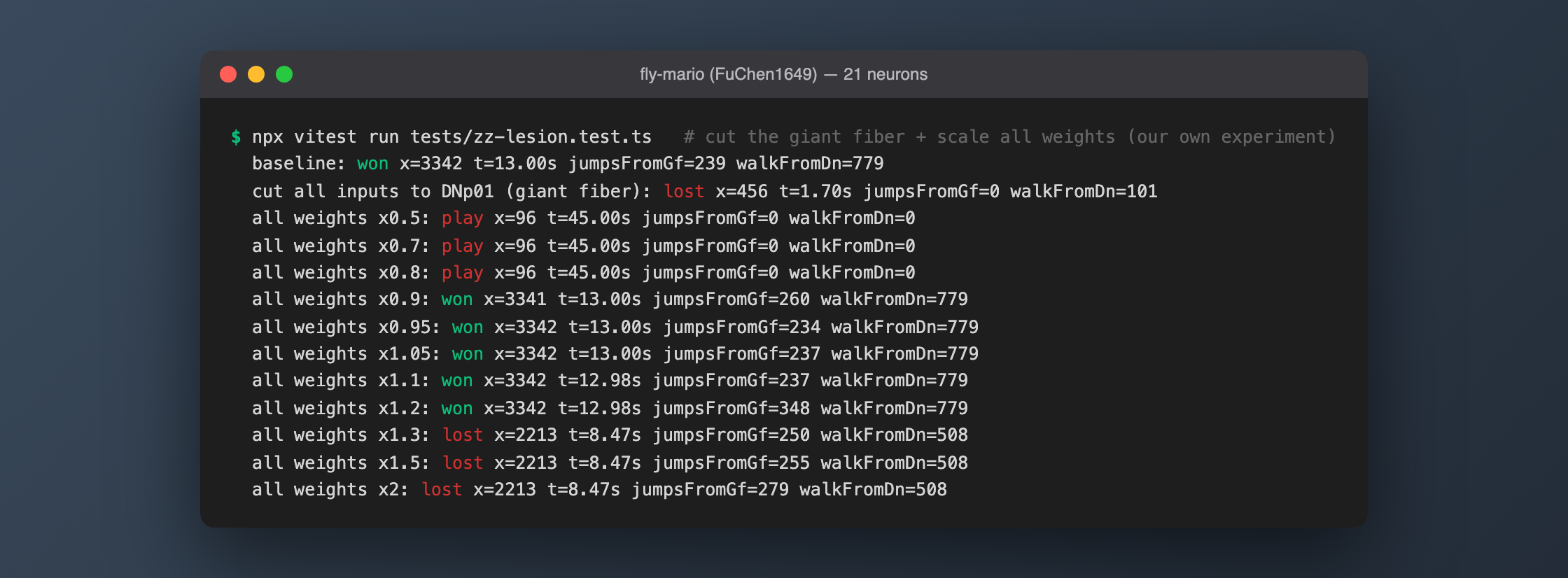
That map has since powered whole-brain simulations, been described as a "fruit fly brain upload", and been wired into Mario and Minecraft by hobbyists. Far fewer people explain how it was made. The answer: one fly, a diamond knife, two custom electron microscopes, a stack of AI, and many years of patience from hundreds of people.
This article goes step by step. The numbers come from the two original papers: Zheng et al. 2018 in Cell (the electron microscopy) and Dorkenwald et al. 2024 in Nature (the FlyWire reconstruction).
The whole process at a glance
| Step | What happened | Key numbers |
|---|---|---|
| ① Choose a fly | One brain picked from several 7-day-old female flies | 1 fly |
| ② Slice | The brain cut into ultra-thin serial sections with a diamond knife | 7,062 sections, 35–40 nm each, about 3 weeks |
| ③ Image | Two custom high-throughput transmission electron microscopes image every section | 21 million images, 106 TB, about 16 months |
| ④ Align | Thousands of section images realigned in 3D | A neural network predicts the deformation between neighbouring sections |
| ⑤ Segment | AI traces every neurite in the images | A convolutional network decides, pixel by pixel, whether it's looking at a cell boundary |
| ⑥ Proofread | People fix the AI's mistakes one by one | 3,013,513 edits, about 33 person-years |
| ⑦ Synapses and labels | Synapses detected, neurotransmitters predicted, cell types annotated | 54.5 million synapses, 8,453 cell types |
①② One fly, 7,062 slices
Why electron microscopy? Because synapses are tiny. The contact between two neurons is tens of nanometres across, while light microscopy is limited by the wavelength of light to about 200 nm. To see every synapse you need electrons.
The catch: electrons can't pass through thick samples, so you can only image very thin sections. The whole brain has to be sliced.
Davi Bock's team at Janelia picked a 7-day-old female fly. Its brain tissue was soaked in heavy metals so cell membranes would show up under the electron beam, embedded in resin, checked with X-ray tomography, and then cut slice by slice with a diamond knife. Each section is 35–40 nm thick — roughly two-thousandths the width of a human hair. The fly brain is about 250 µm deep, so cutting all the way through took 7,062 sections and about three weeks. Three sections went onto each bar-coded metal grid, about 2,400 grids in all.
Four sections were lost during cutting, and 12 in total before full-resolution imaging. 7,050 sections — 99.8% — were imaged successfully.
③ 16 months and 21 million images
Each section is about 0.75 mm wide and 0.35 mm tall: a cross-section of a fly's brain. At 4 nm per pixel, one stitched section is about 187,500 × 87,500 pixels, roughly 16 GB.
A standard electron microscope sees one small field at a time, so the team built two custom high-throughput machines: TEMCA2, with an array of cameras under the microscope shooting simultaneously, and ATPS, a robotic system that picks, aligns and re-stows grids by itself so imaging can run unattended for days.
Acquisition took about 16 months and roughly 2,666 microscopist hours, producing about 21 million camera images, around 106 TB.
The dataset was released publicly in 2018 as FAFB (Full Adult Fly Brain), and everything since has been built on it.
④⑤ Align, then let AI trace the neurons
The 7,050 section images don't line up on their own: sections stretch, wrinkle and shift. A team at Princeton realigned the whole stack with a neural network trained to predict the deformation between neighbouring sections.
Then comes the core step, automated segmentation: deciding which neuron every pixel belongs to, then stitching 7,000-plus layers into individual 3D neurons. FlyWire used a convolutional network that judges, pixel by pixel, whether it's on a cell-membrane boundary, then connects the regions those boundaries enclose.
The AI is fast, but makes two kinds of mistakes:
- False merges: two neurons running close together glued into one;
- False splits: one neuron broken into pieces, especially at fine branches and in lower-quality sections.
A single error can attach a connection to the wrong neuron, so people have to fix them.
⑥ Three million human edits: scientists, professionals and gamers
FlyWire went online in 2019. The idea: put the segmented brain on the web and let people fix it directly in the browser — one click to cut a false merge apart, another to reattach a broken branch.
Three groups did the proofreading:
- Fly researchers. Early on, scientists from more than 50 labs in the FlyWire Consortium proofread the neurons they cared about. More than 200 of them made over 100 edits each.
- Professional proofreading teams. Most of the remaining neurons were later finished by full-time teams at Princeton and Cambridge.
- Gamers. The top 100 players of Eyewire — a citizen-science game where players trace neurons in mouse retina — were invited to beta test. They already had trained eyes for electron microscopy. Calling themselves the "Flyers", they went on to write their own cell-type guides and even build FlyWire plugins.
The bill: the public v783 release required 3,013,513 edits, and the paper estimates about 33 person-years of proofreading. For comparison, the earlier hemibrain (a Janelia–Google collaboration covering only part of the central brain) took about 50 person-years. FlyWire did the whole brain with less effort and similar accuracy.
The work isn't over: the data remains open for proofreading, and corrections go into future releases.
⑦ Find synapses, guess transmitters, name the cells
Once the neurons are traced, you still need to know where they touch.
- Synapses: another AI finds presynapse–postsynapse pairs in the images. About 130 million synapses were detected across the brain, 54.5 million of them between proofread neurons. The fly packs 7.4 synapses into every cubic micron; mammalian cortex has fewer than one.
- Neurotransmitters: AI then predicts from each synapse's appearance whether a neuron releases acetylcholine, glutamate, GABA or another transmitter — which decides whether it excites or inhibits its targets. Every plus or minus sign in today's whole-brain simulations comes from this step.
- Cell types: Schlegel et al. grouped the 139,000 neurons into 8,453 cell types, 4,581 of them new, and matched them against the hemibrain.
Those three layers together are what we call the connectome. I downloaded the public connection table and checked: the synapse-count column sums to 54,492,922, matching the paper (walkthrough in How to Download the Fruit Fly Brain).
So what's the "Google fly brain"?
People searching about the fly brain often look for "Google fly brain". Google was deeply involved in fly connectomics, but not quite the way many assume:
| What Google did | When | Relation to FlyWire |
|---|---|---|
| Flood-Filling Networks, an AI designed for segmenting neurons | Proposed 2016 | The method itself |
| Used them to segment the entire FAFB fly brain (FAFB-FFN1), a forty-teravoxel volume | 2019 preprint | Made manual tracing an order of magnitude faster, but FlyWire didn't use this version — Princeton redid the alignment and segmentation |
| Segmented the hemibrain with Janelia | 2020 | A different dataset |
| Neuroglancer, an open-source tool for viewing 3D microscopy in the browser | — | FlyWire's interface is built on it |
| Partner in the male whole-CNS connectome, MaleCNS | 2026 | Another dataset, including the ventral nerve cord |
So "Google mapped the fruit fly brain" isn't accurate. More precisely: Google supplied several key AI and software tools and produced one automated segmentation; the segmentation and proofreading behind the FlyWire map were done mainly by the Princeton team and the wider community.
After the map
- October 2024: FlyWire is formally published in Nature as a package of papers, data version v783.
- June 2026: Janelia, Cambridge, the MRC Laboratory of Molecular Biology and Google Research release MaleCNS v1.0, the complete central nervous system of a male fly — the first to include the ventral nerve cord, home of the leg and wing motor neurons. The paper appeared in Cell in September.
- Next up is the mouse: a mouse brain has about 500 times as many neurons as a fly's, and so far only one cubic millimetre of cortex has been mapped.
Looking back, the hardest part wasn't the imaging but the error correction. Twenty-one million images took 16 months; fixing the AI's mistakes one by one took hundreds of people several years.
FAQ
Does it matter that the fly was female?
The papers don't explain why a female was chosen. The consequence is that FlyWire lacks male-specific circuitry, and it contains only the brain, without the ventral nerve cord. Both gaps are filled by MaleCNS, the male whole-CNS connectome released in 2026.
It's one fly's brain — does it represent all flies?
Broadly, but they're less "standard parts" than you might think. Schlegel et al. compared FlyWire with the hemibrain from another fly: nearly every hemibrain neuron has a morphological match in FlyWire, and neuron counts and connectivity are broadly stereotyped with occasional variability — yet about a third of the cell types originally proposed for the hemibrain couldn't be reliably re-identified in FlyWire. So they redefined a cell type: cells that are each more similar to cells in a different brain than to any other cell in their own brain.
Can members of the public take part?
FlyWire is still open for proofreading, but editing rights require training and review. To just look, you can browse any neuron's shape and connections on Codex (codex.flywire.ai).
What can I do with this map on my own computer?
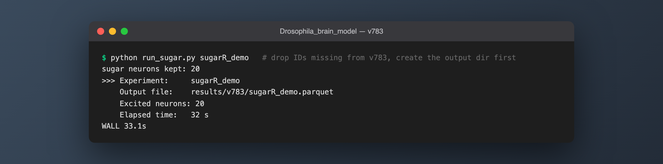
Download it and run the whole-brain model. The official model runs on an ordinary computer; the sugar-taste example finishes in 33 seconds, see How to Download the Fruit Fly Brain and Run It. Or start by driving a physics-simulated fly in your browser at the Digital Fruit Fly Lab.
References
- Zheng et al. 2018, A Complete Electron Microscopy Volume of the Brain of Adult Drosophila melanogaster, Cell. doi:10.1016/j.cell.2018.06.019
- Dorkenwald et al. 2024, Neuronal wiring diagram of an adult brain, Nature. doi:10.1038/s41586-024-07558-y
- Schlegel et al. 2024, Whole-brain annotation and multi-connectome cell typing of Drosophila, Nature. doi:10.1038/s41586-024-07686-5
- Li et al. 2019, Automated Reconstruction of a Serial-Section EM Drosophila Brain with Flood-Filling Networks and Local Realignment, bioRxiv. doi:10.1101/605634
- Januszewski et al. 2018, High-precision automated reconstruction of neurons with flood-filling networks, Nature Methods
- MaleCNS: male-cns.janelia.org